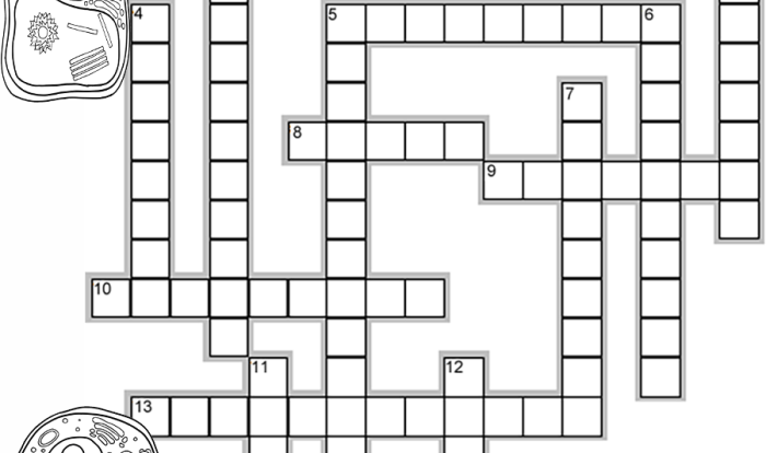
Unlabeled diagram of animal cell – Embark on a captivating journey into the intricate world of animal cells with our unlabeled diagram. This visual masterpiece unveils the essential components and functions of these remarkable building blocks of life, inviting you to explore the depths of cellular biology.
Unveiling the secrets of animal cells, our diagram presents a comprehensive overview of their structure and organization, revealing the significance of unlabeled diagrams in educational settings.
Key Features of an Unlabeled Animal Cell Diagram: Unlabeled Diagram Of Animal Cell
An unlabeled animal cell diagram presents a simplified representation of the cell’s internal structure, omitting labels for specific organelles or components. This type of diagram is commonly used in educational settings to facilitate the study of cell biology and promote active learning.
Purpose of Using Unlabeled Diagrams in Educational Settings
Unlabeled diagrams offer several pedagogical advantages:
- Encourages Observation and Analysis:By withholding labels, unlabeled diagrams require students to carefully observe and analyze the diagram, identifying and interpreting the different structures and their relationships.
- Promotes Active Learning:Students actively engage with the material by labeling the diagram themselves, reinforcing their understanding of the cell’s components and their functions.
- Supports Independent Study:Unlabeled diagrams can be used for self-study and review, allowing students to test their knowledge and identify areas where they need further clarification.
Essential Components of an Animal Cell
Animal cells are the basic unit of life for animals. They are composed of various essential components that work together to perform different functions necessary for the cell’s survival and proper functioning.
The following table provides an overview of the key essential components of an animal cell, along with their functions:
| Component | Description | Function |
|---|---|---|
| Nucleus | The control center of the cell, containing the cell’s DNA. | Directs cell activities, stores genetic information, and regulates gene expression. |
| Cytoplasm | The jelly-like substance that fills the cell, excluding the nucleus. | Provides a medium for cellular activities, contains organelles, and facilitates the movement of substances. |
| Mitochondria | The “powerhouses” of the cell, responsible for energy production. | Generate energy through cellular respiration, producing ATP. |
| Endoplasmic Reticulum (ER) | A network of membranes that extends throughout the cytoplasm. | Involved in protein synthesis, lipid metabolism, and detoxification. |
| Golgi Apparatus | A stack of flattened membranes responsible for processing and packaging proteins. | Modifies, sorts, and packages proteins for secretion or storage. |
| Lysosomes | Membrane-bound vesicles containing digestive enzymes. | Break down and recycle cellular waste, aiding in digestion and cell renewal. |
| Peroxisomes | Small, membrane-bound organelles containing enzymes that detoxify harmful substances. | Metabolize toxic substances, including reactive oxygen species (ROS). |
| Ribosomes | Small, protein-synthesizing structures found in the cytoplasm or attached to the ER. | Translate mRNA into proteins, facilitating protein synthesis. |
| Cytosol | The fluid-filled portion of the cytoplasm, excluding organelles. | Contains dissolved substances, ions, and proteins, and provides a medium for cellular reactions. |
| Cell Membrane | The outermost boundary of the cell, regulating the passage of substances. | Protects the cell, maintains its shape, and controls the exchange of materials with the surrounding environment. |
Comparison of Plant and Animal Cells
Plant and animal cells are both eukaryotic cells, meaning they have a nucleus and other membrane-bound organelles. However, there are also some key differences between these two types of cells.
One of the most obvious differences is that plant cells have a cell wall, while animal cells do not. The cell wall is a rigid structure that surrounds the cell membrane and helps to protect the cell. Plant cells also have chloroplasts, which are organelles that contain chlorophyll and are used for photosynthesis.
Animal cells do not have chloroplasts.
Another difference between plant and animal cells is that plant cells have a large central vacuole, while animal cells have many small vacuoles. The central vacuole is filled with water and helps to maintain the cell’s shape. Animal cells do not have a central vacuole, but they do have many small vacuoles that perform a variety of functions.
Finally, plant cells have centrioles, while animal cells do not. Centrioles are organelles that are involved in cell division. Animal cells do not have centrioles, but they do have a similar structure called the centrosome.
Table: Key Differences Between Plant and Animal Cells
The following table summarizes the key differences between plant and animal cells:
| Feature | Plant Cell | Animal Cell |
|---|---|---|
| Cell wall | Present | Absent |
| Chloroplasts | Present | Absent |
| Central vacuole | Present | Absent |
| Centrioles | Present | Absent |
Functions of Animal Cell Components
Animal cells have various specialized structures called organelles, each with specific functions that contribute to the overall functioning of the cell. Here are some key functions of essential animal cell components:
Nucleus
The nucleus is the control center of the cell, housing the genetic material (DNA). It regulates cell division, protein synthesis, and other cellular activities.
Cytoplasm
The cytoplasm is the jelly-like substance that fills the cell and surrounds the nucleus. It contains various organelles and molecules essential for cell function, such as proteins, enzymes, and nutrients.
Mitochondria
Mitochondria are known as the “powerhouses of the cell” because they generate most of the cell’s energy through a process called cellular respiration.
Ribosomes
Ribosomes are small structures that assemble proteins based on the instructions carried by messenger RNA (mRNA). They can be found freely in the cytoplasm or attached to the endoplasmic reticulum.
Golgi Apparatus
The Golgi apparatus is responsible for processing, modifying, and sorting proteins and lipids. It plays a crucial role in the secretion of cellular products.
Lysosomes, Unlabeled diagram of animal cell
Lysosomes are membrane-bound organelles that contain digestive enzymes. They break down waste materials, recycle cellular components, and defend against invading microorganisms.
Endoplasmic Reticulum
The endoplasmic reticulum is a network of membranes that folds and transports proteins and lipids. It consists of two types: rough endoplasmic reticulum (with ribosomes attached) and smooth endoplasmic reticulum (without ribosomes).
Applications of Animal Cell Diagrams
Unlabeled animal cell diagrams serve as valuable tools in various fields, including education, research, and medical diagnostics. These diagrams provide a simplified representation of the intricate structures and components within an animal cell, facilitating understanding and analysis.
Education
In educational settings, unlabeled animal cell diagrams are widely used as teaching aids. They help students visualize the basic structure and organization of an animal cell. By labeling and identifying the different components, students can gain a deeper understanding of cell biology and its relevance to living organisms.
Research
Animal cell diagrams play a crucial role in research endeavors. Scientists utilize these diagrams to represent and analyze experimental data related to cell structure and function. By comparing and contrasting labeled diagrams of different cell types or under varying conditions, researchers can identify changes or variations in cellular components, providing insights into cellular processes and mechanisms.
Medical Diagnostics
In the field of medical diagnostics, unlabeled animal cell diagrams are employed to assist in the interpretation of microscopy images. Pathologists and other medical professionals use these diagrams as a reference to identify and diagnose abnormalities in cell structure and morphology.
This aids in the diagnosis of various diseases and conditions, such as cancer and genetic disorders.
Top FAQs
What is the purpose of using unlabeled animal cell diagrams?
Unlabeled diagrams facilitate student engagement and active learning by allowing them to identify and label cell components independently, enhancing their understanding of cell structure and function.
How can unlabeled animal cell diagrams be used in education?
In educational settings, unlabeled diagrams serve as valuable tools for interactive learning, quizzes, and assessments, enabling students to demonstrate their knowledge of cell biology.
What are the key differences between plant and animal cells?
Plant cells possess unique features such as a cell wall, chloroplasts, and a large central vacuole, while animal cells lack these structures but have centrioles.